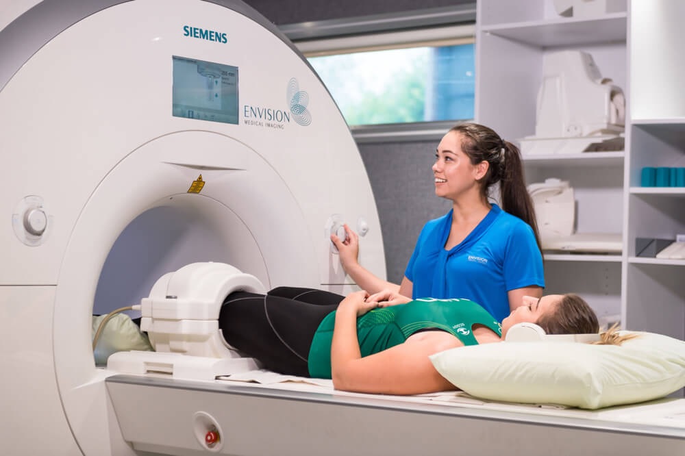
Medical imaging refers to the techniques and processes used to create images of the human body (or parts thereof) for various clinical purposes such as medical procedures and diagnosis.
Diagnostic imaging has become a cornerstone of modern healthcare and continues to improve our quality of life by enhancing the diagnosis of diseases, increasing survival rates, reducing the need for invasive procedures, and furthering our understanding of the human body
Since its inception in the late 1800s and introduction into healthcare in the early 1900s, the field of radiology has evolved from basic plain film X-ray to advanced diagnostic imaging modalities that create three-dimensional cross sections of internal structures.
Each method encompasses specialised techniques designed for the imaging of certain body systems and provides its own set of risks and benefits
The first and oldest type of imaging uses x-rays with the radiation source located outside the patient.
There are five primary x-ray imaging modalities and these are:
– Plain film x-ray which is the oldest form of medical imaging;
– Fluoroscopy which is the continuous acquisition of a sequence of X-ray images over time;
– Computed Tomography; the use of X-ray beams from various angles to produce cross-sectional images referred to as slices that may be reconstructed into 2D or 3D volumes of anatomic structures;
– Dual-emission x-ray absorptiometry (DXA) which uses two different energies of ionising radiation to measure a patient’s bone mineral density; and
– Mammography which uses low energy x-rays to specifically image breast tissues.
A second type of medical. imaging commonly referred to as nuclear medicine produces medical images by viewing a radioactive chemical or compound that has been introduced into the patient’s body.
Nuclear medicine encompasses three main modalities and they are Nuclear Medicine Planar Imaging, Positron Emission Tomography (PET) & Single Photon Emission Computed Tomography (SPECT).
Finally and not least, imaging modalities that do not use ionising radiation include Ultrasound which is constructed by projecting high frequency sound waves into the body and using sonic echoes to create a real-time image of internal anatomy & Magnetic Resonance Imaging (MRI) which uses magnetic fields and radio frequency pulses to create 2D and 3D images with amazing clarity.
While the various forms of imaging technology provide us with the tools to image anatomy and detect diseases in ways previously only imagined, there may be a great risk of over-radiation that can result from the inappropriate or unnecessary use of these procedures.
The decision to order diagnostic imaging is mostly always driven by weighing the potential benefit against the potential risk.
When an imaging study is considered beneficial, the quality of the image and the care taken to perform the study correctly are always taken into consideration.
The overall approach to radiation safety followed by any personnel working in medical imaging is and should always be the concept of “as low as reasonably achievable,” or ALARA, with the goal of all imaging procedures being to keep the radiation dose as low as possible while providing a high-quality diagnostic image.
Though medical/clinical judgment may be sufficient prior to treatment of many conditions, the use of diagnostic imaging services is paramount in confirming, correctly assessing and documenting courses of many diseases, as well as in assessing responses to treatment.
This week we celebrate World Radiography Day and ISRRT has chosen this year’s theme as “Elevating Patient Care with Artificial Intelligence”.
Artificial intelligence and machine learning have enough potential to make various tasks in the healthcare industry possible with accurate performance.
Patient’s timely disease diagnosis and the right decision is an important part of hospitals to improve the overall healthcare system.
Artificial intelligence is going to play a big role in diagnosing the various types of diseases including the critical maladies with a high level of accuracy.
The Radiology Department at Oceania Hospital has been around for close to two decades and has been moving in line with the continuing evolution in medical imaging science from Digital X-ray units, 3D/4D Ultrasound to the recent introduction of its 256 slice GE Revolution HD Computed Tomography and the World Class Innova IGS 530 in the Catheterization Lab. As medical imaging progresses with innovation, OHPL Radiology Department will continue to introduce new & improved technologies to improve healthcare.
As we celebrate World Radiography Day, we celebrate the discovery of X-rays by Wilhelm Roentgen in 1895 & it’s evolution throughout the years since its inception in the healthcare facility.
We celebrate the crucial role medical imaging plays in the field of diagnostics & most importantly, we celebrate us, personnel that work with radiation and/or medical imaging as a whole.
As professionals, we make a difference because we understand that the cornerstone of the principle of radiation protection for our patients is Justification and Optimization.
Happy World Radiography Day!
* Maria Vatunilagi is a senior radiographer / sonographer at Oceania Hospitals Pte Ltd. The views expressed are the author’s and not necessarily shared by this newspaper.


